70 year male was bought to Casuality in an intubated state
HOPI:
Pt is apparently asymptomatic until 2 days ago then he developed sudden onset of abdominal discomfort and SOB after dinner. Patient was taken to nearby hospital in altered sensorium and was found Grbs-30 mg/dl . patient was intubated i/v/o poor GCS-5/15 and was bought to our hospital for further management with ET tube in situ
PAST HISTORY:
H/O Right inguinal surgery on 15/09/23
K/c/o hypertension since 12 yrs( on unknown medication)
K/c/o Pulmonary Tuberculosis 30 years back( used ATT for 6 months)
PERSONAL HISTORY:
DIET :mixed
APPETITE : Normal
SLEEP: adequate
BOWEL AND BLADDER :Regular
Addictions : Patient was an Alcoholic and smoker 30 years ; stopped after diagnosed with Tuberculosis.
FAMILY HISTORY:
N/K/C/O DM, Hypertension,Epilepsy, Asthma, Thyroid disorders.
GENERAL EXAMINATION:
Patient is on Mechanical ventilation.
Dilated neck veins present.
No Pallor,Icterus,clubbing,cynosis.
VITALS:
TEMP: 97.2 F
BP: 160/90 MM/HG
PR: 98 BPM
GRBS: 91mg/dl
SYSTEMIC EXAMINATION:
RS:
Position of trachea :deviated to right,
Movement of chest decreased on right side.
B/L rhonchi at IAA,ISA.
B/L Crepts at MA,AA,IAA,ISA.
CVS : S1 , S2 heard, no precordial bulge, apical impulse at left 6th ICS 1cm medial to MCL.
P/A :distended abdomen,no organomegaly,bowel sounds -sluggish
CNS: GCS :E1VtM1
Tone - normal in all four limbs
Reflexes- absent
Plantars- mute

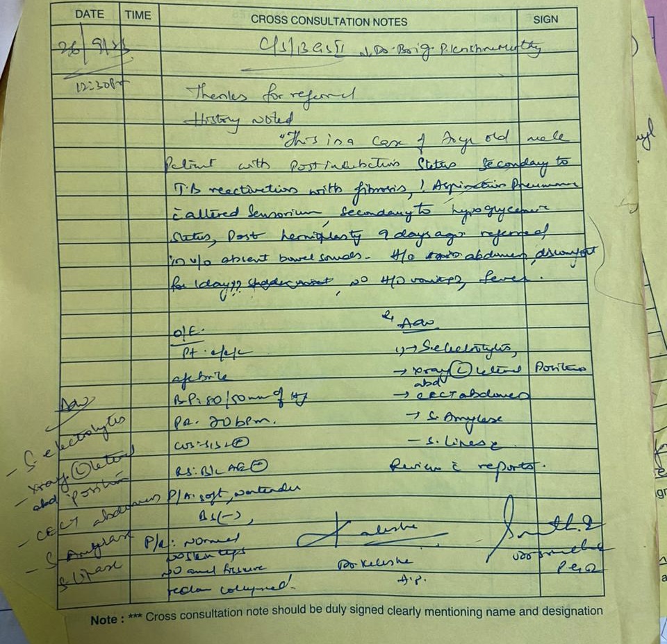
PROVISIONAL DIAGNOSIS
ALTERED SENSORIUM SECONDARY TO ?HYPOGLYCAEMIA
?ASPIRATIONAL PNEUMONIA
POST INTUBATION STATUS
S/P RIGHT HERNIOPLASTY
INVESTIGATIONS
CHEST X-RAY
24-09-2023

26-09-2023


|
Hemogram |
25/9/23 |
26/9/23 |
|
Hb |
9.4 |
10 |
|
TLC |
18400 |
19500 |
|
PCV |
31.8 |
35.2 |
|
MCV |
83.2 |
85 |
|
MCH |
24.6 |
24.2 |
|
MCHC |
29.6 |
28.4 |
|
RDW-CV |
17.7 |
17.6 |
|
RBC COUNT |
3.82 |
4.14 |
|
PLATELET |
1.89 |
1.57 |
|
SMEAR |
NC-HC |
NC-HC |
|
N/L/E/M/B |
84/05/01/10/00 |
83/02/00/15/00 |
|
LFT |
24/9/23 |
26/9/23 |
27/9/23 |
|
TB |
0.47 |
0.82 |
0.80 |
|
DB |
0.16 |
0.15 |
0.19 |
|
AST |
13 |
15 |
220 |
|
ALT |
18 |
8 |
83 |
|
ALP |
100 |
123 |
129 |
|
TP |
6.1 |
6.2 |
5.2 |
|
ALB |
3.3 |
3.1 |
2.01 |
|
A/G |
1.18 |
1.03 |
0.63 |
|
RFT |
24/9/23 |
26/9/23 |
|
UREA |
20 |
31 |
|
CREATININE |
0.8 |
0.8 |
|
URIC ACID |
2.4 |
2.9 |
|
CALCIUM |
9.3 |
9.9 |
|
PHOSPHOROUS |
4.1 |
3.1 |
|
SODIUM |
137 |
137 |
|
POTASSIUM |
4.3 |
4.4 |
|
CHLORIDE |
102 |
103 |
|
ABG |
24/9/23 |
25/9/23 |
26/9/23 |
|
|
5:59AM |
11PM |
|||
|
pH |
7.19 |
7.17 |
7.23 |
6.71 |
|
pCO2 |
80.1 |
63.2 |
70.8 |
220 |
|
pO2 |
130 |
80.5 |
101 |
82 |
|
HCO3 |
29.4 |
22.4 |
28.7 |
26.7 |
|
St.HCO3 |
24.2 |
19.8 |
24.8 |
12.5 |
|
BEB |
-0.3 |
-5.5 |
0.4 |
-15.4 |
|
BEECF |
1.9 |
-4.9 |
1.8 |
-9.2 |
|
TCO2 |
63.2 |
50.5 |
61.8 |
70.6 |
|
O2 Sat |
97.2 |
95.6 |
97.1 |
83.2 |
|
O2 count |
15.9 |
10.1 |
14.1 |
12.2 |
|
|
25/9/23 |
|
BT |
2.30sec |
|
CT |
5sec |
|
APTT |
34sec |
|
PT |
17sec |
|
INR |
1.25sec |
|
|
24/9/23 |
|
Anti-HCV |
Non reactive |
|
HBSAG |
Negative |
|
HIV |
Non reactive |
|
Blood Group |
A+ve |
|
Blood Lactate |
9 |
|
RBS |
90 |
|
|
26/9/23 |
|
Serum lipase |
22 |
|
Serum amylase |
40.8 |
|
LDH |
225 |
|
|
27/9/23 |
|
Pleural sugar |
79 |
|
Pleural protein |
2.2 |
|
Pleural amylase |
10 |
|
Pleural LDH |
210 |
24-09-2023
Pulmonology referal was taken in view of chest X-ray changes


TREATMENT
1) RT FEEDS - 100ml water 2nd hourly, 200ml milk+ protein powder 4th hourly
2) IV FLUID NS @50ml/hr
3) INJ MIDAZOLAM + FENTANYL @4ml/hr increase/decrease accordingly
4) INJ PIPTAZ 4.5gm IV/BD
5) INJ CLINDAMYCIN 600mg IV/TID
6) INJ PAN 40 mg IV/OD
7) INJ ZOFER 4mg IV/TID
8) NEBULISATION WITH DUOLIN 8th HOURLY
9) NEBULISATION WITH MUCOMIST 12th HOURLY
10) NEBULISATION WITH IPRAVENT 4th HOURLY
11)ET TUBE SUCTIONING 2nd HOURLY
12)POSISTION CHANGE 2nd HOURLY

TREATMENT
1) RT FEEDS - 100ml water 2nd hourly, 200ml milk+ protein powder 4th hourly
2) IV FLUID NS @50ml/hr
3) INJ MIDAZOLAM + FENTANYL @4ml/hr increase/decrease accordingly
4) INJ VANCOMYCIN 1gm IV/BD
5) INJ CEFTAZIDIME 2gm IV/TID
6) INJ CLINDAMYCIN 600mg IV/TID
7) INJ PAN 40 mg IV/OD
8) INJ ZOFER 4mg IV/TID
9) NEBULISATION WITH DUOLIN 8th HOURLY
10) NEBULISATION WITH MUCOMIST 12th HOURLY
11) NEBULISATION WITH IPRAVENT 4th HOURLY
12)ET TUBE SUCTIONING 2nd HOURLY
13)POSISTION CHANGE 2nd HOURLY
14) TAB ISONIAZID 5mg/kg 350mg
TAB RIFAMPICIN 10mg/kg 700mg
TAB PYRIZINAMIDE 25mg/kg 1750
TAB ETHAMBUTOL 15mg/kg 1050
DIAGNOSIS
POST INTUBATION STATUS SECONDARY TO ASPIRATION PNEUMONIA WITH ALTERED SENSORIUM SECONDARY TO HYPOGLYCEMIA(GCS 5/15) WITH RIGHT LUNG BRONCHECTIASIS WITH RIGHT LUNG FIBROSIS SECONDARY TO POST TUBERCULAR SEQUELAE(30 YEARS AGO) WITH S/P HERNIOPLASTY(RIGHT INGUINAL)POD-10 WITH K/CIO
HT SINCE 12 YEARS
DEATH SUMMARY
70 YEAR MALE WHO WAS A K/C/O PULMONARY TB WITH POST TB FIBROSIS OF RIGHT LUNG AND HT SINCE 12 YEARS WITH A HISTORY RIGHT INGUINAL HERNIOPLASTY POD-8 WAS BROUGHT TO ER(24/9/23 5:20PM) WITH ET TUBE INSITU ON MECHANICAL VENTILATION ACMV-VC MODE UPON ADMISSION ABG SHOWED TYPE 2 RESP FAILURE, RESP ACIDOSIS.
NECESSARY INVESTIGATIONS WERE DONE AND PATIENT WAS SHIFTED TO ICU.
PULMONARY REFERRAL WAS TAKEN AND ADVISE FOLLOWED. HRCT SHOWED CONSOLIDATION IN MULTIPLE SEGMENT IN LEFT LUNG LOWER LOBE-ACUTE INFECTION.
BRONCHECTIASIS IN ANTERIOR SEGMENT OF LEFT LUNG UPPER LOBE AND SEQUELEA OF CHRONIC INFECTION. PULMONOLOGY REVIEW REFERRAL WAS TAKEN AND ADVISE FOLLOWED.
SURGERY REFFERAL WAS TAKEN I//O NO BOWEL SOUNDS AND ADVISE FOLLOWED.
SERIAL ABGS SHOWED PROGRESION OF RESPIRATORY ACIDOSIS. IN/O INCREAING PCO2 GRADUAL FALL IN SATURATION PATIENT HAS BEEN SHIFTED TO ACMV-VC MODE TO ACMV-PC MODE. DESPITE ALL THE EFFORTS PATIENT HAD FALL IN SATURATIONS AND BP. PATIENT WAS STARTED ON IONOTROPES IN/O FALL IN SATURATION; ABSENT CENTRAL AND PERIPHERAL PULSES PATIENT WAS INITIATED ON CPR.PATIENT COUNT BE REVIVED AND DECLARED DEATH ON 26/9/23 11:21PM

Comments
Post a Comment